Symptoms of breast cancer include a lump in the breast, bloody discharge from the nipple and changes in the shape or texture of the nipple or breast. Redness, swollen lymph nodes, discomfort or thickening or puckering of the skin are also experienced by some people.
Get the answer of all your queries regarding IVF from our fertility specialist. To book an appointment Call 78980–47572 / 80852–77666 or For more detail visit www.mohakivf.com
Best infertility hospital in indore, ivf specialist in indore, ivf center in indore, test tube baby center in indore, ivf treatment cost in indore, Best centre for IVF in indore, affordable ivf cost in indore, best fertility hospital in india, best ivf center in mp, infertility treatment in indore, best ivf centre in india, ICSI treatment in indore
The purpose of this study was the analysis of a correlation, in infertile patients, between the quality of the endometrium based on its vascularisation and the chances of conception. Hysteroscopy was carried out to determine the quality of the endometrial surface using the Sakumoto–Masamoto classification (“good” vs. “poor” endometrium) in the secretory phase of the menstrual cycle. The results were set in relation to the outcome of the subsequent infertility treatment, i.e. the establishment of a pregnancy within the study period (4 years). In 108 (67%) of the 162 followed-up patients, the endometrium was endoscopically classified as “good”, while in 54 (33%) the result was “poor”. The overall pregnancy rate was 37% (60 patients); 47 of all pregnancies (78%) occurred in women with a “good” endometrium while 13 (22%) had a “poor” classification. This positive association between the establishment of a pregnancy in the follow-up and a “good” classification of the endometrial vasculature in the group with a “good” endometrium was significant (P = 0.0165, Fisher’s exact test). This study confirms the usefulness of endometrial evaluation by hysteroscopy as a diagnostic instrument for providing a prognosis of the chance for the patients to become pregnant.
Background
One of the most difficult questions put forward by patients after the failure of a fertility therapy such as in vitro fertilisation (IVF) and intra-cytoplasmic sperm injection (ICSI) is related to the lack of success. The implantation rate per transferred embryo normally does not exceed 30%. Often the failure of “embryo implantation” is given as an explanation as the failure in one of the most critical stages at the beginning of conception, i.e. when apposition and implantation has to occur inside the uterine cavity. Current knowledge about the mechanism of these interactions is still difficult to interpret [1].
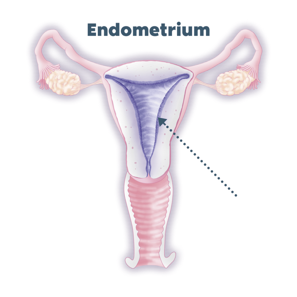
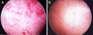
Various different suggestions have been made for investigating these mechanisms and attempting to understand which would be the characteristic elements of the endometrium that ensure ideal conditions for the embryo; but they have until today been limited to the so-called theory of the endometrial “opportunity window” [2] and did not offer effective clinical instruments for understanding which groups of patients would be at an increased risk of embryo implantation failure [3]. By using hysteroscopy as a diagnostic procedure for the assessment of pathologies inside the uterine cavity, it has, however, been shown that the differential characterisation of the endometrial surface could be a helpful tool for evaluating the in vivo vascularisation of the uterine mucosa. Already, Sakumoto et al. in 1992 in the first place [4], and after him Masamoto et al. in 2000 [5], have described the technique and used this differentiation in order to demonstrate that the endometrium could be classified into two distinct groups: a “good” endometrium, which has circular gland openings and an intense vascular ramification on one hand, and a “poor” endometrium, which is characterised by a surface with a lower gland and vascular density on the other.
The purpose of this study was to demonstrate the impact of the hysteroscopy, according to this vascularisation-based staging, and to investigate whether this endometrium quality could be used as a tool to assess the potential to achieve a pregnancy irrespective of the chosen type of infertility treatment.
Materials and methods
All infertile patients attending our fertility centre and with a regular menstrual cycle were asked to participate in this comparative, prospective study. They underwent a pre-operative transvaginal sonography (TVS), a full hormonal assessment (FSH, LH, 17β-estradiol, thyroid-stimulating hormone and prolactin) in the serum on cycle days 3 to 5 and then a hysteroscopy in the second part of the menstrual cycle for evaluating the vascularisation of the endometrium. Informed, written consent was obtained from the patients after explanation of the study by the clinician prior to the procedure, and they were asked to avoid a pregnancy in the examination cycle. The study protocol was approved by the local ethical committee.
The inclusion criteria were infertility (absence of conception after 12 months of regular, unprotected intercourse), age less than 43 years, regular cycles (25–31 days) and normal hormonal values (including FSH <12 mU/mL) had to be fulfilled. All partners provided a spermiogram for the exclusion of male factor infertility. Further exclusion criteria were known causes of uterine malformations, endometrial adhesions and hormonal therapy such as oral contraceptives or other oestrogen–progesterone medications within the last 3 months before hysteroscopy. If necessary, the procedure was combined with a laparoscopy to test the tubal patency, and the hysteroscopy was done in most cases during the same operating session and under general anaesthesia. The ultrasonographers were located in the same university department, but not involved in the surgical procedure, and the surgeon was blinded to the TVS findings.
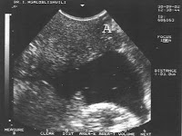
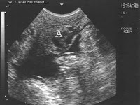
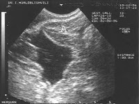
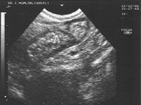
The endometrial surface was evaluated according to the Sakumoto–Masamoto grading (“good” vs. “poor”). Endoscopic findings were categorised as “good” with an appearance representing ring-type glandular openings and maximal glandular secretion or “poor” with a low development level of vessel networks on the endometrial surface. This is illustrated in Fig.1. Hysteroscopic procedures were carried out when indicated (e.g. polyps, myomas, adhesions, septa). The diagnostic hysteroscopy was performed with a 5-mm-outer diameter scope (30°, Karl Storz) connected to a standard endoscopic camera, and a saline solution at low pressure (not higher than 60 mmHg) was used for the distension of the uterine cavity. Hysteroscopic findings were observed and analysed by three gynaecologists using videotape records.
The follow-up interval lasted for 12 months from hysteroscopy. Data were recorded and analysed for a correlation between the vascularisation score of the endometrium and the occurrence of embryo implantation (spontaneous pregnancy, successful outcome after hormonal stimulation with or without intrauterine insemination or successful IVF/ICSI-embryo transfer treatment). For statistical evaluation, the Fisher’s exact test was applied using GraphPad Prism Software (San Diego, USA). For alpha, we considered 0.05 as cutoff value to avoid type I error.
Findings
A total of 178 infertile women underwent a hysteroscopic assessment, and 162 (91%) of them could be followed up in our hospital. A “good” endometrium according to Sakumoto–Masamoto staging was diagnosed in 108 of them (67%), while 54 (33%) patients were graded as “poor”. No differences in the distribution pattern of the causes and duration of infertility, the age of the patients (mean 33.8 years in the “good” and 33.6 in the “poor” group) or the pre-treatment day 3 serum level of follicle-stimulating hormone (6.8 and 7.4 U/L) were observed between these two groups.
A normal uterine cavity was reported in 133 (83%) women, while endometrial polyps, submucosal fibroids, adhesions or uterine malformations were found in 29 cases (17%). On the other hand, the pre-operative TVS indicated intrauterine pathologies in 15 cases (9.3%). The overall pregnancy rate was 37% (60 women); 15 women became pregnant spontaneously, 22 patients succeeded after follicular stimulation with recombinant gonadotropins (rFSH) and 23 after treatment with in vitro fertilisation and embryo transfer including ICSI.
In the total pregnancy group (N = 60), a “good” endometrium was found in 47 women (78%) while this was the case in 61 patients (60%) of the group who did not achieve a pregnancy. Forty-one patients with a “poor” endometrium did not succeed in getting pregnant. Only 13 patients with a “poor” endometrium did succeed in establishing pregnancy in the follow-up. The association between endometrium quality by Sakumoto–Masamoto classification and pregnancy outcome was statistically significant (P = 0.0165, OR = 2.43, CI = 1.17–5.05); the contingency matrix for the pregnancy outcome is shown in Table 1.
Conclusion
Our results confirm those of the studies carried out by Sakumoto and Masamoto [4, 5], indicating that a hysteroscopic examination of the mid-secretory endometrium can be a reliable instrument for determining the chances of a patient to become pregnant. The classification in “good” and “poor” is leading to the conclusion that a poorly vascularised endometrium with limited glandular (secretory) structures may result in a tissue which is not suitable for a correct embryo implantation and endometrial development, and this irrespective of other factors of sterility.
Nevertheless, our results showed a lower fraction of patients (one third) with a “poor” endometrium in comparison to earlier studies (45.9% in the study of Sakumoto [4] and 61.3% in Masamoto et al. [5]): we believe that this difference can be explained with a different patient selection in the study groups. As a matter of fact, we did not focus on patients with a history of repeated abortions as it was the case in the study of Masamoto [5], but on a global infertile population.
Another clearly interesting but only partially surprising finding is the high percentage (17.2%) of intrauterine pathologies that have been diagnosed in the hysteroscopic examination when compared to the total number of patients with suspected intracavitary problems found in the pre-operative sonography (9.3% of all women, and this in spite of all ultrasound examinations having been carried out by the same team of experienced gynaecologists). These results, nevertheless, are in large agreement with previously published studies [6, 7].
We therefore conclude that a hysteroscopic examination, particularly in cases of idiopathic infertility or after several unsuccessful treatment cycles with in vitro fertilisation [8], is strongly indicated [9] and has the added benefit of providing a prognostic measure for determining the chances of the patient to become pregnant, in the future, in addition to its diagnostic significance [10].
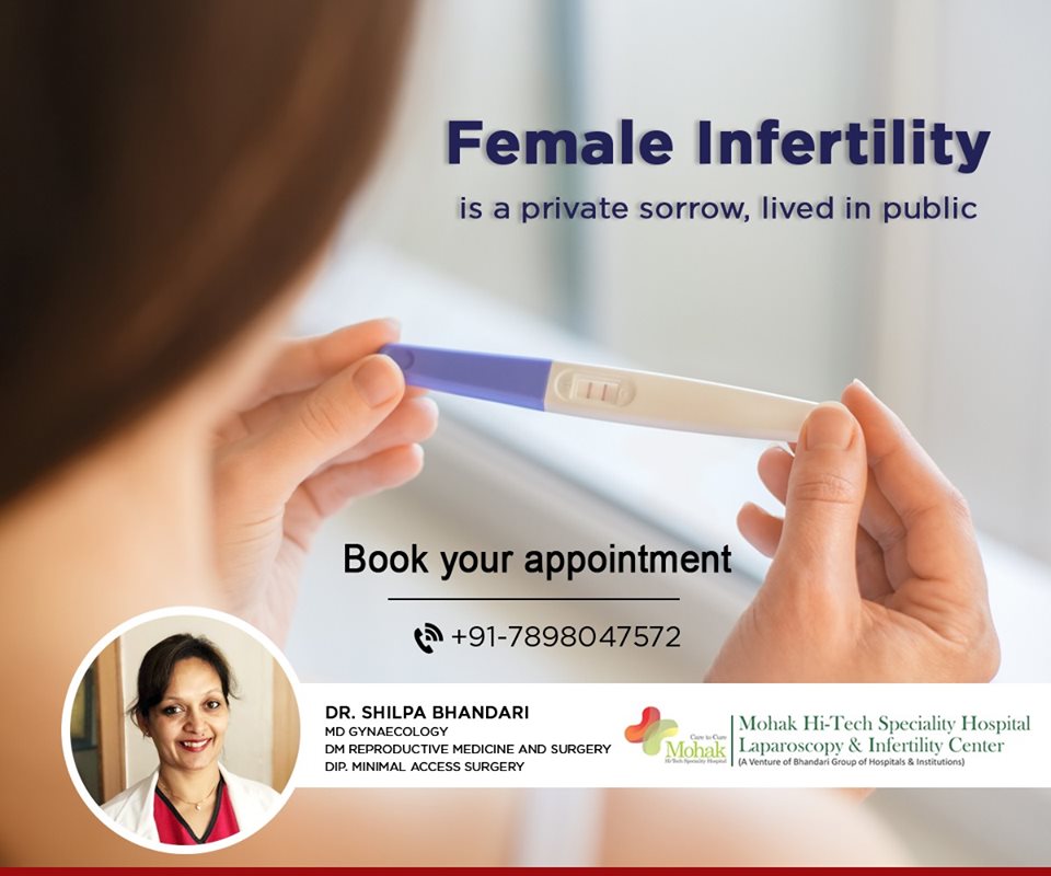
Are you looking for the Best IVF specialist in indore? Dr Shilpa Bhandari is one of the first reproductive medicine specialist of this country. She has not only been a pioneering force in the development of an MCI recognized DM/ McH reproductive medicine curriculum, but has also procured this prestigious super specialization by 3 years of extensive training in this field.
She looks after the needs of more than a thousand patients annually in terms of consultation, surgeries, IVF, medical treatment, etc. she has also been actively involved in training other doctors as well as research. She has authored more than 20 research papers in national and international journals. Dr Shilpa Bhandari is an ardent believer in open patient communication, maintaining and honest doctor patient relationship and patient empowerment. Her dream is to provide affordable, honest patient care to couple seeking to enhance their families. Book an appointment Today Call now 7898047572 For more information, visit – https://www.mohakivf.com
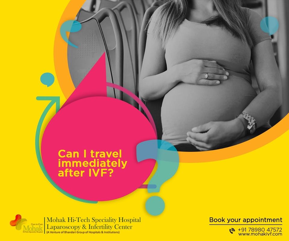
There is no reason for a complete restriction on physical activity during IVF. For many women, exercise is a stress relief strategy. Undergoing IVF is a naturally stressful process, and it is important to use all of your resources to reduce anxiety and tension. However, you should keep your exercise routine to a low-impact maximum during the cycle. While mild to moderate exercise is acceptable during IVF treatment, patients should be careful about what types of exercises they perform. Focus on low-impact exercises that encourage stress relief.
It’s also important to drink plenty of water before, during and after a workout, and to eat a healthy and well-balanced diet to keep your energy up. Above all, remember: Taking care of your body should be your top priority during your IVF cycle.
Get the answer of all your queries regarding IVF from our fertility specialist. To book an appointment Call 78980–47572 / 80852–77666 or For more detail visit www.mohakivf.com
Best infertility hospital in indore, ivf specialist in indore, ivf center in indore, test tube baby center in indore, ivf treatment cost in indore, Best centre for IVF in indore, affordable ivf cost in indore, best fertility hospital in india, best ivf center in mp, infertility treatment in indore, best ivf centre in india, ICSI treatment in indore
Hypothalamic Dysfunction Excess Physical or emotional stress, a very high or a recent substantial weight gain or loss can disrupt production of certain hormones and affect ovulation.
Get the answer of all your queries regarding IVF from our fertility specialist. To book an appointment Call 78980–47572 / 80852–77666 or For more detail visit www.mohakivf.com
One of the intensively discussed topics recently is investigation of infertile patients. A subject of particular interest is a new technique – transvaginal hydrolaparoscopy (TVHL), which has already found its place amongst other methods of infertility exploration. Transvaginal hydrolaparoscopy allows atraumatic investigation of reproductive organs in physiologic conditions, in ambulatory settings. Significant, for clinical practice, is the possibility of a combination of transvaginal hydrolaparoscopy with other methods of investigation – sonohysterosalpinogography, hysteroscopy and salpingoscopy, which make possible integrated exploration of the reproductive system. Instillation of saline into the pelvic cavity through uterine tubes (or through puncture needle if needed) under ultrasonographic control and exposition of posterior fornix, allows better orientation in the pouch of Douglas, complex assessment of reproductive organs and determination of advisability of pelvioscopy – the relatively invasive technique as well as thorough control of the safe access to the pouch of Douglas.
Since Gordts et al. in 1998 have introduced the technique of transvaginal hydrolaparoscopy (TVHL), for the investigation of infertile women [1], interest to the vaginal route of genital tract exploration has been recommenced.
Introduction of more global concept of fertiloscopy: concurrent performance of pelvioscopy, hysteroscopy, salpingoscopy and microsalpingoscopy with chromopertubation [2] allows for an increase in usefulness of infertility investigation and to optimize treatment strategy for infertility management with the consequent saving of time and costs. Introduction of this highly informative endoscopic technique into the format of one-stop investigation of infertility has become possible after the introduction of a new technique of atraumatic access to the pouch of Douglas via vaginal route using new instrumentations for transvaginal hydrolaparoscopy (TVHL) in the ambulatory settings [3, 4].
From April of 2002 till November of 2005 we have performed 837 fertiloscopies. As we described previously [5], TVHL with hysteroscopy and salpingoscopy is routinely performed in our clinic, in complex with sonohysterosalpingography and with extensive use of ultrasonographic control, in order to assess the future diagnostic insertion into the pouch of Douglas, as well as to ensure safety of this manipulation (instillation of warm saline for exposition of the posterior fornix of vagina).
The most difficult stage of TVHL is the introduction of instruments through the posterior fornix of the vagina into the pouch of Douglas. According to the data of various authors, access to the pouch of Douglas failed in 4.3-0% cases [1–4, 6–15]. Complications are expected when the patient has a retroverted uterus or in the presence of ovaries, uterine tubes, leiomyoma, bowel loops or varicose veins in the pouch of Douglas. Some authors consider such disposition as a relative/absolute contraindication to the procedure [1, 3, 4, 7, 12, 14, 16]. According to the data, the bowel trauma took place in 0.65% of procedures, but after the initial experience it has reduced to 0.25[16]. In 50% of cases, bowel injury was estimated by the surgeon as avoidable. The development of safe techniques for insertion of instrument/s into the pelvic cavity, as well as determination of the capability and the necessity of performing pelvioscopy is highly important.
As a matter of fact, nowadays there are several, similar systems of instruments for the access to the pouch of Douglas which differ in construction of the puncture needle (combined needle-trocar system with adapted Veress needle, and separate needle and trocar system). All systems have their advantages and disadvantages, but discussion of these topics is not the aim of this article. We would like to introduce some safe methods which enable the avoidance of complications to a grater extent, and to specify the possibility and rationality (reasonableness) of the performance of TVHL.
During performance of procedures we use Transvaginal hydrolaparoscopic kit (Circon ACMI, Stamford, CT) or Transvaginal hydrolaparoscopy set (Karl Storz, Tuttlingen, Germany). Ultrasonographic investigation/assistance was performed using Aloca SSD 650 with transvaginal probe with a frequency of 5.5 MHz, equipped with the puncture set.
During the process of preparation for laparoscopic investigation, detailed ultrasonography is performed with particular attention to the location of uterine corpus, uterine cervix and vagina, also to the state of posterior fornix of the vagina and the contents of the pouch of Douglas.
The first stage of the investigation is routine hysteroscopy, after which the pouch of Douglas is partially filled with saline (if at least one of the uterine tubes is patent).
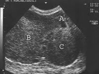
The second stage is the routine sonohysterosalpingography during which by transtubal instillation of 60–200 ml of saline into the pelvic cavity, exposition of the posterior fornix of vagina is reached, which makes it possible, against a background of instilled fluid, to clearly visualize and assess the fornix (Figs. 1, 2, 3 and 4 determine the presence, the location and the characteristics of adhesions in the pouch of Douglas, presence of dilated vessels and presenting organs in minor pelvis. (Figs. 5, 6, 7 and 8).
In the case of non-patency of uterine tubes, which makes the instillation of saline into the pelvic cavity impossible, transvaginal puncture set is used under the ultrasonographic control, to allow exposition of the posterior fornix by instillation of water via an aspiration needle Fig. 7. The presence of follicular fluid during ovulation sometimes is quite enough for ultrasonographic orientation in the situation.
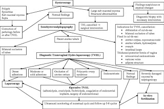
The third stage of investigation is the transvaginal hydrolaparoscopy: an insertion of laparascopic instruments into the pelvic cavity is performed, if there have not been revealed contraindications or reasons of TVHL cancellation (for example hydrosalpinges-Fig. 8) at previous stages of investigation. These conditions are:
Complete obliteration of the pouch of Douglas Fig. 5.
Thickening of posterior fornix, often with uneven echo structure and width Fig. 2, with the visualized dilated vessels Fig. 3, sometimes with presence of retro cervical endometriosis or varicose veins, adipose structure behind the posterior fornix or vagina Fig. 4.
Intensive adhesions in the pouch of Douglas Fig. 7.
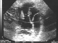
Localization of fixed organs of minor pelvis into the pouch of Douglas: one or both ovaries, uterine tube/s Fig. 8, intestinal loops Fig. 5, myomatous node or uterine corpus Fig. 6.
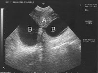
Bilateral hydrosalpinges Bilateral Fig. 8.
Among the investigated 837 infertile women, in 6 cases was detected suspicious pathology on atypical alterations, thus further stages of investigation were postponed until an appropriate differential diagnosis is obtained.
Results of the next stage, sonohysterosalpingography for the remaining 831 women are summarized in Table.
After the stages of hysteroscopy and sonohysterosalpingography, transvaginal hydrolaparoscopy was performed in 702 of the 837 women. An extent of exploration was determined during the procedure, after considering advisability and possible safety of conducting different interventions (Fig. 9 Algorithm of management). In 135 (16.1%) women out of 837 TVHL was cancelled or postponed. We have not noted any complications during performed investigations.
Hydrosalpinges of at least one of the tubes, does not represent contraindication for performing transvaginal hydrolaparoscopy per se (if it is not obliterating the pouch of Douglas), but often on the stage of sonohysterosalpingography/hystreoscopy the need for the transvaginal hydrolaparoscopy vanishes, as a treatment strategy of infertility becomes evident (Fig. 9 Algorithm of management). On the basis of already discovered findings it is possible to switch to the accepted interventions for infertility management. This includes either restoration of natural fertility – reconstructive surgery of the uterine tubes or creation of optimal conditions for IVF (proximal obstruction of tubes, salpingectomy etc) [17].
Ultrasonographic control during procedure of transvaginal hydrolaparoscopy not only allows an evaluation of the proximal parts of tubes which in complex increases informativeness of the investigation, but also gives a possibility to visualize existence of adhesions in the pouch of Douglas-together with the introduction of the trocar system. However it is not sufficient enough to assess the role of adhesions in infertility as this method is not able to access the fimbrial-ovarian area- a key functioning part of the reproductive tract.
Thus, we present the investigation of infertility which includes: hysteroscopy, sonohysterosalpingography with instillation of fluid into the pelvic cavity (if necessary via puncture needle) under ultrasonographic control, which provides exposition of the posterior fornix and orientation in the pouch of Douglas, hydropelvioscopy with dye chromopertubation and salpingoscopy. Such a format of investigation makes it possible for amore complex evaluation of reproductive system and determination of advisability of performance of transvaginal hydrolaparoscopy – a relatively invasive procedure with more intensive control of safety of the access to the pouch of Douglas.
Are you looking for the Best Fertility hospital in India? Mohak Infertility Center is one of the leading Best Fertility hospitals and Test tube baby center in indore Mohak Infertility Center is a IVF center in Indore Madhya Pradesh with an excellent and experienced team of IVF specialist and embryologists, who are able to offer you today’s most advanced and effective procedures in IVF treatment. Our IVF Specialists and embryologists have been trained and have worked in some of the Best IVF Centers of the world, Mohak Infertility Center in Indore aims to give you the best chance of having a healthy baby. Our team of IVF specialist and support staff is committed to provide you the best possible care and advise that you can trust. Book an appointment Today Call now 7898047572 For more information, visit – https://www.mohakivf.com
Feeling Emotions and Overthinking — mental health playing important role in reproductive hormone regulation if we are stressed it may affect to conceive a baby.
Get the answer of all your queries regarding IVF from our fertility specialist. To book an appointment Call 78980–47572 / 80852–77666 or For more detail visit www.mohakivf.com
Get the answer of all your queries regarding IVF from our fertility specialist. To book an appointment Call 78980–47572 / 80852–77666 or For more detail visit www.mohakivf.com
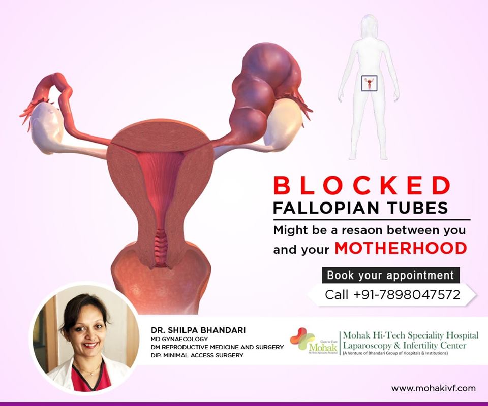
Tubal factor infertility accounts for approximately 25–35% of cases of female infertility [1–3]. Identifiable causes of tubal infertility are postinfectious tubal damage, post surgical adhesion formation, and endometriosis-related adhesions.
The normal process of captation of the oocyte requires a series of prerequisites: the ovarian surface free from adhesions, the fimbrial-ampullary portion of the tube free to embrace the ovary and, beside tubal patency, a normal activity of the ciliated and secretory cells of the tubal mucosa. Furthermore, the muscular layer of the tube must be undamaged and able to contract.
In recent years the treatment of tubal infertility has witnessed a shift from tubal reconstructive surgery to in vitro fertilization–embryo transfer techniques (IVF). Due to the wider availability of assisted reproductive technologies, the number of women with mechanical infertility treated by reconstructive surgery has decreased, most couples being referred to IVF.
Reproductive surgery is performed with the aim of allowing ovum pick-up by restoring normal anatomic relationships between the fimbriae and the ovary. However, even though reproductive surgery may be successful in restoring normal anatomy, it may not be able to restore normal function of the damaged tubal mucosa.
The percentages of success of the surgical treatment are therefore strictly correlated with the type of preexisting tubal damage, independently of the surgical technique performed.
Recent refinements of laparoscopic instrumentation and increased surgical skills in operative laparoscopy allow laparotomy to be avoided in most instances.
The advent of salpingoscopy, a technique that allows direct visual evaluation of the tubal mucosa, has allowed improved selection of patients who are candidates for tubal reconstructive surgery by identifying the patients with good reproductive prognosis. The following is an analysis of the various indications to tubal surgery according to the level (proximal or distal) and type of tubal pathology.
Proximal tubal occlusion (PTO) Lack of passage of the contrast medium at the level of the intramural–isthmic portion of the fallopian tube during an hysterosalpingogram (HSG) or a laparoscopy with chromopertubation may be due to a true occlusion consequent to postinfectious fibrosis or to an obstruction due to technical artifacts, a spasm of the uterine tubal ostium, a valve mechanism determined by an area of endometrial thickness (focal hyperplasia), or to plugs of amorphous material.
Bilateral PTO is a relatively infrequent finding. We reported [4] that out of 665 patients undergoing laparoscopy with chromopertubation for primary or secondary infertility, only 35 patients (5%) had bilateral PTO confirming a previous HSG finding (25 patients bilateral, 10 unilateral with the contralateral tube either distally occluded or absent). Of these patients, 17 refused any further treatment. After a mean follow-up of 25 months, 3 (18%) of these patients spontaneously conceived an intrauterine pregnancy; 4 out of 5 patients who underwent a repeated HSG had bilateral tubal patency. Therefore, the diagnosis of bilateral tubal occlusion proved to be incorrect in 7 out of 17 patients (42%).
Furthermore, with regard to the etiology of temporary proximal tubal obstruction, a recent paper [5] hypothesizes that small air bubbles, but more likely tubal kinking, may be an explanation of these findings in the patients undergoing HSG in the supine position. In a series of 156 patients, unilateral PTO was diagnosed in 15% of patients (24 of 156) and bilateral PTO in 3% (4 of 156). Rotating the patient such that the obstructed tube was inferior to the uterus resolved 63% of the unilateral PTO, likely by unkinking the tube at the uterotubal junction, thus dramatically lowering the resistance to the flow of contrast medium. The same manoeuvre was less effective in bilateral PTO, where 25% of the more dependent tubes became patent. Still, this report offers an important contribution to the explanation of “reversible” PTO. The possibility that some PTO are obstructions and not true occlusions is supported by the study of Sulak et al. [6] who in 1987 reported on 18 patients who were found to have bilateral PTO by both HSG and subsequent laparoscopy with chromopertubation and therefore underwent resection of the occluded tubal segment and anastomosis. Resected tubal segments were studied histologically, and in 11 of the 18 cases no tubal occlusion could be demonstrated. In six cases (three with occlusion and three with apparent patency) the tubal lumen contained an amorphous material of unknown etiology, often appearing to form a cast of the tube. The authors were the first to report on such “plugs” and speculated that, if they cause tubal obstruction, this would explain previously published findings of high pregnancy rates in infertility patients after HSG. The suggested mechanism would be, among others, dislodging of tubal mucus plugs.
In 1987 Thurmond et al. [7] described their technique for selective salpingography and fallopian tube recanalization that has since then been widely used to improve diagnosis by injecting contrast medium through a catheter placed in the tubal ostium. This technique allows differentiation of tubal spasm from true occlusion, and can be performed in the same session as the hysterosalpingographic examination that fails to opacify the tubes. In fallopian tube recanalization, a catheter and guide wire system is used to clear proximal tubal obstruction by amorphous debris.
A review [8] evaluating results with this technique in 1,466 patients reports a successful recanalization of the proximal fallopian tube in 71–92% of recanalization attempted. Pregnancy rates after the procedure have been variable among series, with an average rate of 30% during follow-up.
In a retrospective study, Al-Jaroudi [9] et al. have recently evaluated the reproductive performance of women after selective tubal catheterization. Ninety-eight infertile women with hysterosalpingographic findings of PTO underwent a repeat hysterosalpingography examination before selective tubal catheterization. Bilateral tubal patency was documented in 14 patients and patency of one of the tubes in 12 others. PTO was confirmed in 72 patients. Successful recanalization of both tubes was achieved in 25 patients (34.7%) and successful recanalization of at least one tube was achieved in 44 patients (61.1%). Of the 72 patients who underwent selective tubal catheterization, 23 conceived (31.9%). The cumulative probability of conception was 28%, 59%, and 73% at 12, 18, and 24 months of follow-up, respectively. The few patients with failure of tubal recanalization may likely have true occlusion caused by fibrotic scarring of the tube from salpingitis, endometriosis, or surgery. Microsurgical resection and tubocornual anastomosis continue to be the standard of care in these cases [10]. In a review of nine case series including 187 patients with PTO, we reported [11] a 49% term pregnancy rate per patient, with a 4% risk of ectopic pregnancy after microsurgery by laparotomy.
In 1987, Patton et al. [12] reported on a series of 27 patients with a postpelvic inflammatory disease (PID) bilateral PTO or PTO of the single remaining tube diagnosed both at HSG and laparoscopy with tubal perfusion. Patients were not excluded on the basis of age, extent of tubal disease, duration of infertility, tubal length, or history of prior operation. After an extended follow-up (mean 1,714 days) the possibility of conception was of 46%, 65%, and 69.3% within 1, 2 and 3 years from surgery, respectively.
The probability of a conception resulting in a live birth was 27%, 47%, and 53.2% at 1, 2, and 3 years after surgery, respectively. When only patients who did not have a previous surgery for infertility were considered, the conception rate was 75% with a live birth rate of 58% after 3 years.
Periadnexal adhesions In case of periadnexal adhesions, the classic open-abdomen surgery has been completely replaced by laparoscopic surgery, because it obtains the same results in terms of reproductive outcomes with all the advantages of the laparoscopic approach (better cosmetic result, minor postoperative pain, shorter hospital stay, decreased risk of postoperative infections, quicker return to work).
The intrauterine pregnancy rate reported in the literature after laparoscopic salpingoovariolysis in nonselected patients ranges from 51–62%, and the ectopic pregnancy rate ranges from 5–8% [10].
Recent prospective studies have demonstrated that the most important prognostic factor in terms of reproductive outcome after reproductive surgery is the status of the tubal mucosa as evaluated by salpingoscopy [13–16]. For salpingoscopy, a 2.8-mm rigid salpingoscope that allows a detailed vision of the tubal ampullary mucosa is used. The salpingoscope is introduced into the abdominal cavity through the operating channel of the laparoscope. The abdominal ostium of the tube is identified and cannulated, and the tubal mucosa is evaluated. At the ampullary level four or five major folds are noted, with minor folds interspersed between them.
The status of the tubal mucosa is classified according to the classification proposed by Brosens et al. [13] as follows: grade I—normal mucosal folds are seen; grade II—the major folds are separated and flattened but otherwise normal (might be considered a grade I distended by increased intraluminal hydrostatic pressure); grade III—focal adhesions are seen between the mucosal folds; grade IV—extensive adhesions are present between the mucosal folds and/or disseminated flat areas are noticed; grade V—there is a complete loss of the mucosal fold pattern.
Grades I and II identify a normal mucosa; grades III to V identify a tubal damaged by a previous pelvic infectious disease.
It has to be stressed that there is no correlation between the score of periadnexal adhesions and intraluminal damage.
Salpingoscopy allows the identification of the patients with normal tubal mucosa who may most benefit from laparoscopic salpingoovariolysis, with a term pregnancy rate of 70%. Data from Brosens and Marana [14–16] indicate that about 80% of patients with periadnexal adhesions have a normal tubal mucosa. Therefore, 80% of the patients with periadnexal adhesions have a normal mucosa, with 70% chance of a term pregnancy after a laparoscopic salpingoovariolysis. Most of the pregnancies occur within 1 year of surgery.
Distal tubal occlusion (DTO) Salpingoneostomy utilizing microsurgical techniques, first described by Swolin [17] in 1967, has been for years the procedure for the treatment of distal tubal occlusion. In a literature review of 14 series, including 1,275 patients, we reported [18] a cumulative intrauterine pregnancy rate with microsurgical salpingoneostomy by laparotomy of 326/1275 (26%). The cumulative term pregnancy rate was 239/1158 (21%), the cumulative spontaneous abortion rate 54/1125 (5%), and the cumulative ectopic pregnancy rate 96/1245 (8%). Ten studies, including 1,128 patients, had complete information on pregnancy outcomes. The cumulative pregnancy rate per patient was 371/1128 (33%). Of the pregnancies, 77% (284/371) were intrauterine, 61% (227/371) were term pregnancies 15% (55/371) were spontaneous abortions, and 23% (87/371) were ectopic pregnancies.
A recent review evaluated five nonrandomized control studies that compared laparoscopic and open microsurgical tubal surgery for treatment of DTO [19]. No significant difference was observed in the intrauterine pregnancy rate between the two groups (laparotomy group: 138/478, 28.9%; laparoscopy group: 104/336, 30.9%; combined OR 1.32 [95% CI 0.58–3.02]). In three of the studies, sufficient information was given to compare surgical techniques used at different stages of tubal disease.
Overall, there was no significant difference in the intrauterine pregnancy rate in laparatomy versus laparoscopy in mild tubal disease (laparotomy group: 83/253, 32.8%; laparoscopy group: 96/243, 39.5% OR 1.06 [95% CI 0.42–2.70]).
For patients with severe stage tubal disease, there was a significantly increased intrauterine pregnancy rate in the laparotomy group (47/210, 22.4% versus 6/86, 6.98%, OR 2.88 [95% CI 1.16– 7.16]).
Subsequently, the principles of microsurgery were introduced in the laparoscopic approach for the treatment of distal tubal disease.
Several classifications have been proposed in order to identify the patients that may most benefit from tubal reproductive surgery in DTO. Various parameters are considered, such as the type and extension of periadnexal adhesions, the degree of tubal occlusion, and the status of the tubal mucosa.
In 1988, the American Fertility Society proposed a scoring system in order to allow the comparison of results obtained from different authors. This was based on the following parameters: type and extension of the adhesions and, in addition, for the classification of distal tubal occlusion, thickness and rigidity of the tubal wall, distal ampullary diameter, and the percentage of mucosal folds preserved at the neostomy site. The importance of intraoperative salpingoscopy to visualize the entire length of the ampullary mucosa was recognized. However, salpingoscopic findings were not included in the scoring system as salpingoscopy was being practiced in very few centers.
Numerous prospective studies have recently demonstrated that, also in the case of distal tubal occlusion, the most important prognostic factor is represented by the status of the tubal mucosa. It is therefore important to identify the patients with normal tubal mucosa by means of salpingoscopy.
In fact, prospective studies have demonstrated that patients with normal tubal mucosa (grades I and II) will have a term pregnancy rate of 65% after salpingoneostomy (compared to 25% obtained in nonselected patients).
Studies of Brosens and Marana [14–16] report that in cases of distal tubal occlusion, the percentage of patients with normal tubal mucosa range from 35–45%. Therefore, in cases of DTO, 35–45% of the patients have a normal tubal mucosa, with a 65% chance of a term pregnancy rate after a laparoscopic salpingoneostomy. Most of the pregnancies occur in 12–18 months.
In conclusion, based on these findings, in cases of DTO, our current approach would be a diagnostic laparoscopy with salpingoscopy. Laparoscopic salpingoneostomy would then be performed in the patients with normal tubal mucosa.
Reversal of tubal sterilization Tubal sterilization is one of the most used contraceptive methods around the world. It has been reported that about 1% of the patients undergoing this procedure subsequently request a reversal of tubal sterilization.
Tubo-tubal anastomosis is best performed with microsurgical techniques by laparotomy. The precision afforded by this procedure allows precise excision of the occluded segments and exact approximation of each layer of the proximal and distal portions of the tube.
As in the majority of cases the tubal segments are normal, the outcome is an anatomically and physiologically normal tube although slightly shorter.
This leads to a high intrauterine pregnancy rate with a low risk of ectopic pregnancy. Gomel and McComb [10] have reported a cumulative intrauterine pregnancy rate of 70% in patients who are <35 years of age and a 55% rate in patients who are 35 years of age or more at the time of reversal, with most pregnancies occurring within 18 months after surgery. The ectopic pregnancy rate is approximately 2%.
Recent improvements in laparoscopic microsurgical instrumentation have prompted a few centers to propose tubal anastomosis by laparoscopic access. In a retrospective clinical study, Yoon et al. [20] reported on 202 women who desired reversal of tubal sterilization. In these patients tubal anastomosis was performed by laparoscopy. The cumulative pregnancy rate in the 186 patients for whom follow-up data were available was 60.3%, 79.4%, and 83.3% at 6, 12, and 18 months after surgery, respectively. Five patients (3.2%) had ectopic pregnancies; one of these patients subsequently conceived an intrauterine pregnancy.
The authors concluded that laparoscopic tubal anastomosis is less invasive and could be an alternative to the procedure by laparotomy.
Gomel and McComb [10] contend that the mechanical disadvantages inherent in laparoscopic surgery will lead inevitably to less precision than that readily attainable by microsurgery by minilaparatomy for any given surgeon. At present there are no randomized trials with sufficient number of patients to answer this question. IVF results According to the American Society for Reproductive Medicine/Society for Assisted Reproductive Technology Registry published in 2007 [21], reporting the results of 79,042 IVF cycles (with and without ICSI) performed in 2001, the percentage of clinical pregnancy was 32.8% per initiated cycle, 38.2% per retrieval, and 40.6% per transfer. The delivery rates were, respectively, 27.2%, 31.6%, and 33.6%. The cancellation rate was 14.1%; the clinical pregnancy loss was 17.2% and the ectopic pregnancy rate 1.8%.
Of the deliveries, 64.1% were singletons, 32.0% were twins, 3.7% were triplets, and 0.1% were greater than triplet deliveries.
According to the European Society of Human Reproduction and Embriology Registry published in 2007 [22], reporting the results of 365,000 ART cycles performed in 2003, the clinical pregnancy rate per retrieval and per transfer were, respectively, 26.1% and 29.1% for IVF, whereas they were 26.5% and 28.7%, respectively, for ICSI. Incomplete data were available for the analysis per cycle and for term deliveries.
Of the deliveries, 76.7% were singleton, 22.0% were twins, and 1.1% triplets.
The latest results published by the North American and European societies reported here confirm a trend toward better results for assisted reproductive techniques with passing years. The same improvements are not present for the results of tubal surgery. A major improvement with this respect has, however, been made in the field of better patient selection for tubal surgery, following which, for example, as previously discussed, a global 25% pregnancy rate in nonselected patients with DTO can be brought up to 65%. In the final section of this review, a personal view on the comparison between IVF and tubal surgery is reported.
Discussion It is important to underline that while IVF is a ‘palliative’ technique, which means that it does not eliminate the problem but bypasses it, surgery is curative in the favourable cases with normal tubal mucosa. This allows women to obtain pregnancy naturally, and it is therefore an option for couples with ethical and religious concerns. If successful, surgery allows women to have more than one pregnancy without further treatment, with an abortion rate similar to that of the normal population.
Indications to IVF for ‘tubal factor infertility’ may not be correct as this diagnosis often proves to be fallacious. In fact, we have demonstrated that the diagnosis of PTO has a high false positive rate due to technical problems, valve mechanism, intraluminal debris, or chronic inflammation. The diagnosis of DTO, although generally accurate, may sometimes be mimicked by ampullary diverticulae, due to a congenital defect of the myosalpinx, that do not need reconstructive surgery and are not incompatible with pregnancy [23].
In a recent study, Hennelly et al. [24] sent a questionnaire to each patient who was known to have delivered an infant after an IVF or ICSI treatment at their university-based assisted reproduction unit and who had not returned for further therapy. Five hundred fifty questionnaires were sent out. Five hundred thirteen (94%) responses were received and analyzed. One hundred six (20.7%) of the 513 respondents reported that they had had a subsequent spontaneous pregnancy. All the pregnancies occurred within 2 years of the IVF/ICSI pregnancy success. The authors underlined that patients entered the program only if they had a valid indication for IVF/ICSI. These patients truly undertook IVF as a last resort. Therefore, it was surprising to find that 19 out of 128 patients with a diagnosis of tubal factory infertility (14.8%) later conceived spontaneously.
With respect to financial concerns, it should be considered that, unlike in the USA, in Italy as in other European countries, operative laparoscopy, even for infertility, is fully subsidized by the government health service when performed in a public hospital. On the contrary, IVF is mainly performed in private centres and is not reimbursed either by the government or private insurances.
The risks of tubal surgery are very low and are due to the known complications of anesthesia and surgery. Although low, the risk of complications is present even in IVF, with a reported prevalence of serious cases of ovarian hyperstimulation syndrome of 14 per 1,000 women after the first cycle and 23 per 1,000 after a mean of 3.3 treatments in the 9,175 patients followed by the National Research and Development Centre of Finland [25].
With regard to cumulative pregnancy rate after IVF, in a recent paper Sharma et al. [26] reported a cumulative live birth rate of 66% following four cycles of IVF. However, the discontinuation rate was very high during the study. Only 36% of patients continued treatment after the first unsuccessful attempt (dropout rate 74%); the dropout rate was 61% after the second attempt, and 69% after the third attempt.
Lack of success and psychological stress are the main factors in influencing the decision to discontinue treatment with increasing number of attempts [27]. A prospective, cohort study reported that an unexpectedly high percentage of couples who performed IVF discontinued the subsidized treatment before the three cycles that were offered. The majority of these discontinuations were due to psychological stress [28].
Concern has recently been expressed about the health of the children conceived after IVF [29, 30]. It has been reported in singleton ART infants a two-fold increase in risk of perinatal mortality, low birthweight, and preterm birth, about a 50% increase in small for gestational age, and a 30–35% increase in birth defects [31]. The same Centre for Child Health Research evaluated all papers published by March 2003 with data relating to the prevalence of birth defects in infants conceived following IVF/ICSI compared with spontaneously conceived infants [32]. Meta-analyses of seven reviewer-selected studies and of all 25 studies identified as suitable for inclusion in a meta-analysis suggest a statistically significant 30–40% increased risk of birth defects associated with ART. The authors conclude that this information should be made available to couples seeking ART treatment.
The guidelines recently approved by the Genetics Committee and the Reproductive Endocrinology and Infertility Committee of the Society of Obstetricians and Gynecologists of Canada for counselling of Canadian women using ART recommend: pregnancy achieved by IVF with or without ICSI are at higher risk for obstetrical and perinatal complications than spontaneous pregnancies; singleton pregnancies achieved by ART are at higher risk than spontaneous pregnancies for adverse perinatal outcomes, including perinatal mortality, preterm delivery and low birth weight; ART has a significant risk of multiple pregnancies; risks of multiple pregnancies include higher rates of perinatal mortality, preterm birth, low birth weight, gestational hypertension, placental abruption, and placenta previa; and that further epidemiologic and basic science research is needed to help determine the etiology and extent of the increased risks of congenital abnormalities associated with ART [33].
An increased risk of congenital malformations in relation to IVF even in singleton infants has been confirmed by a recent review analysing the medical literature update to 2006 [34]. In conclusion, in spite of the recent improvements in the success of IVF, tubal reconstructive surgery remains an important option for many couples. In referral centers, surgery should be the first line approach for a correct diagnosis and treatment of tubal infertility. The success of the surgical treatment depends on careful selection of patients using appropriate diagnostic techniques.
Are you a childless couple frantically searching the Best IVF center in India with Highest IVF success rate? If yes, Mohak Infertility Center is one of the Best Infertility hospitals and IVF centers in indore. Provides you International Standard Infertility Treatment in indore along with assisted reproductive technologies like IUI, IVF, ICSI ,etc at affordable IVF treatment / Test tube Baby Treatment cost along with the satisfaction of being consulted by the most renowned IVF specialist in Indore. it requires of you. Book an appointment Call now 7898047572 For more information, visit – https://www.mohakivf.com